What are Ankle Cartilage injuries?
The ankle bone, (the talus) can develop tears in the cartilage lining of the joint, which then also form cysts in the bone underneath.
These are termed Osteochondral defects, or an OCD.
This is usually due to an injury such as a sprain. These often cause swelling and pain, and can be associated with catching or sharp intermittent pain in the ankle.
An OCD has a natural history of not healing and progressing to more advanced cartilage and leading to arthritis. A small number may heal with rest and protection.

Treatment options range from non-operative to operative.
NON-OPERATIVE MANAGEMENT
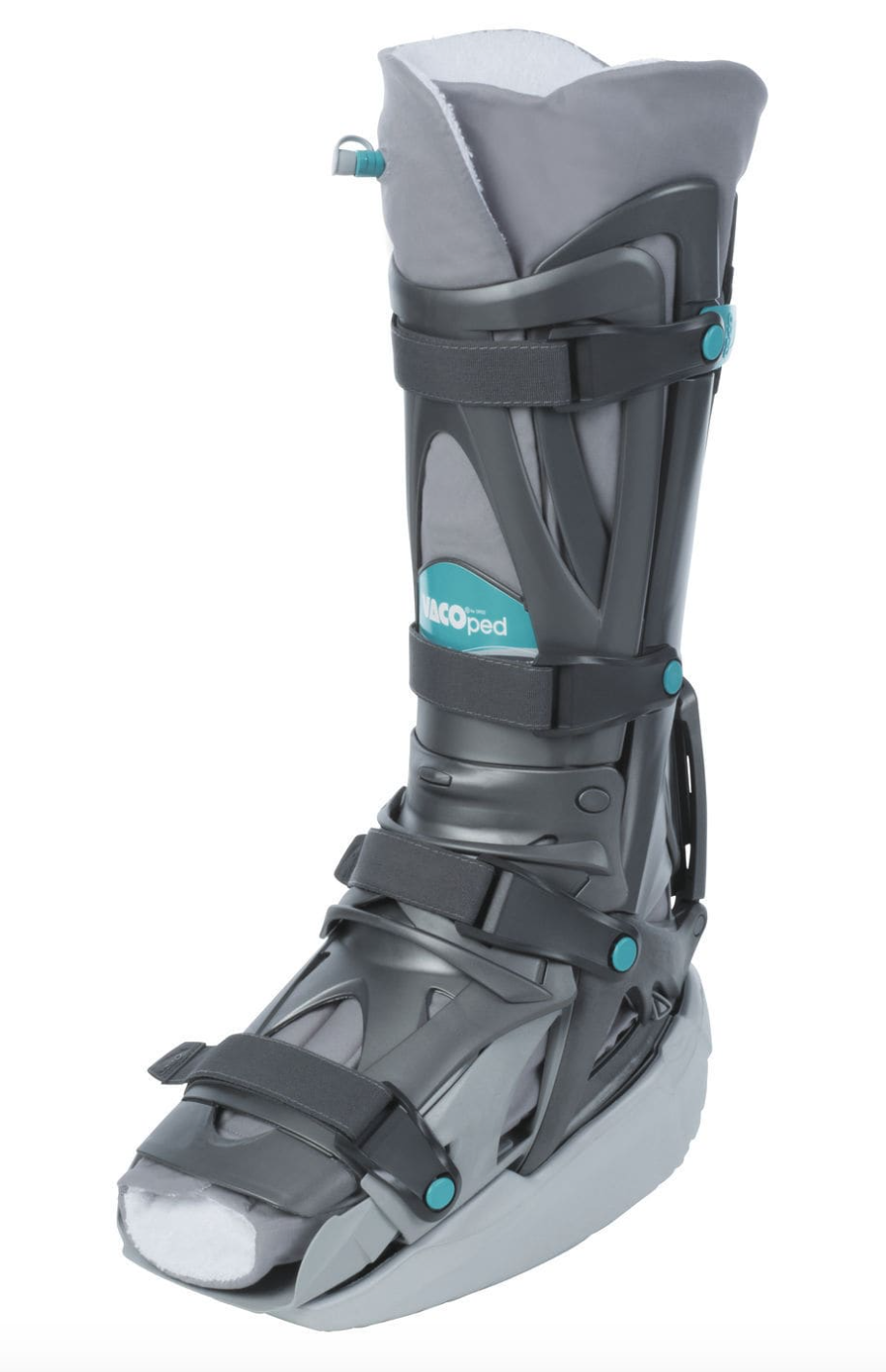
Immobilisation and Protected weight bearing may be effective in early minor cases. A CAM boot and crutches can be used, followed by a strengthening program.
OPERATIVE MANAGEMENT
Arthroscopic treatment is the most common and initial surgical management. The rationale is to remove the frayed and torn cartilage, create a stable rim, and then shave the base to create an even bleeding surface. A synthetic Gel is then injected into lesion, which initially forms a scaffold and promotes the formation of a stable regrowth of a cartilage scar.
Currently evidence suggest this has an up to 85 % chance of healing an OCD. Extensive research over the last 20 years has analysed treatment options such a stem cell transplants and PRP injections. The key component is the physical shaving of the ankle bone to create a stable base.
Following surgery the foot is elevated for 2 weeks. In most cases weight bearing is allowed in a CAM boot. Ankle movement and strengthening can commence after 2 weeks. No impact or high level sport should be performed for up to 3 months.
There are small risks of skin numbness, stiffness, ongoing pain , and recurrence of the condition.
